• Francesco Renzi's Research Group
  • Francesco Renzi's Research Group
  • Francesco Renzi's Research Group
  • Francesco Renzi's Research Group
  • Francesco Renzi's Research Group
  • Francesco Renzi's Research Group
  • Francesco Renzi's Research Group
  • Francesco Renzi's Research Group
  • Francesco Renzi's Research Group
  • Francesco Renzi's Research Group

Research

The surface structures of C. canimorsus

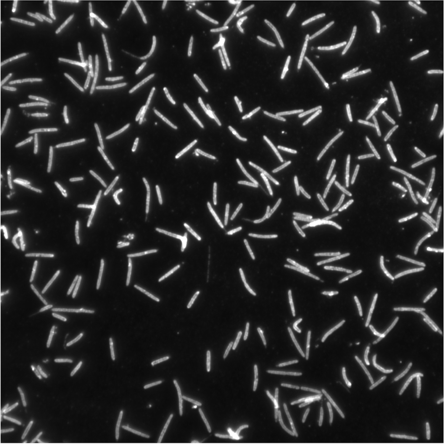
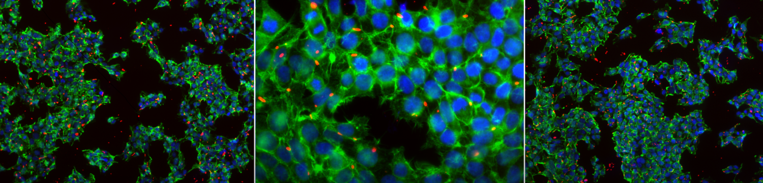
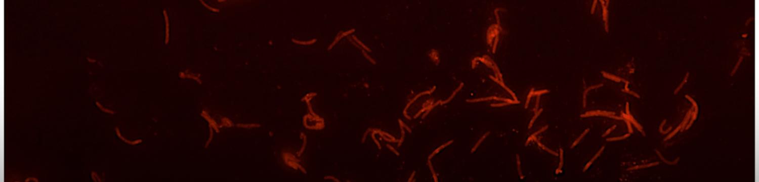
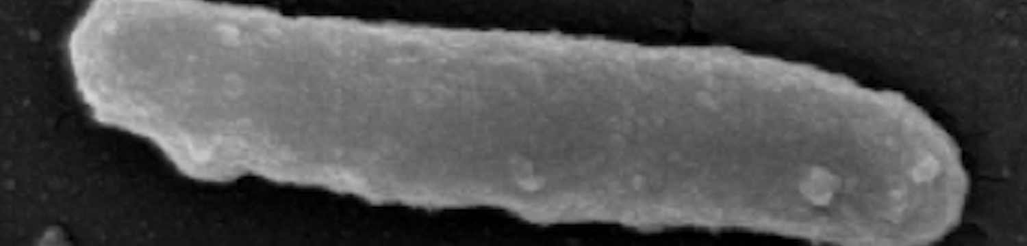
The surface structures of bacteria are essential for their survival in the host and hence their pathogenicity.  They may represent a protection against the attack of complement and against phagocytosis but at the same time, they are an Achille's heel since the innate immunity system has evolved to recognize them to trigger inflammation.  We investigate the structure and role of the different surface structures of Capnocytophaga canimorsus, in collaboration with U. Zähringer (Borstel, D), I. Sadovskaya (Boulogne-sur-mer, F) and E. Maes (Lille, F).


Publications:

- Renzi, F*, Hess, E*, Dol, M, Koudad, D, Carlier, E, Ohlén, M, Moore, E & Cornelis, GR 2018, Capsular serovars of virulent Capnocytophaga canimorsus are shared by the closely related species C. canis and C. cynodegmi  Emerging Microbes & Infections, vol. 7, no. 1, 124, pp. 124. DOI: 10.1038/s41426-018-0126-x * equal contribution.

- Hess, E*, Renzi, F*, Karhunen, P*, Dol, M, Lefevre, A, Antikainen, J, Carlier, E, Hästbacka, J & Cornelis, G 2018, Capnocytophaga canimorsus Capsular Serovar and Disease Severity, Helsinki Hospital District, Finland, 2000–2017 Emerging Infectious Diseases, vol. 24, no. 12, pp. 2195-2201. DOI: 10.3201/eid2412.172060 * equal contribution.

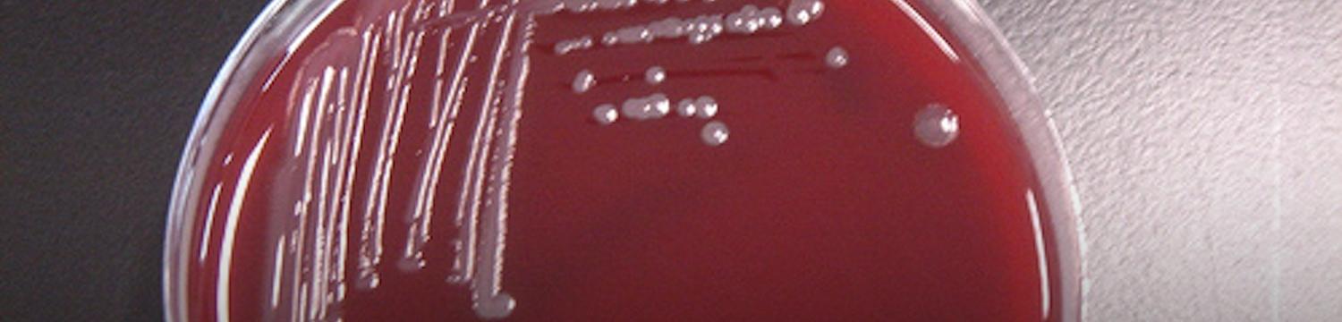
- Hess, E*, Renzi, F*, Koudad, D, Dol, M & Cornelis, G 2017, Identification of virulent Capnocytophaga canimorsus isolates by capsular typing Journal of clinical microbiology, vol. 55, no. 6, pp. 1902-1914. DOI: 10.1128/JCM.00249-17 * equal contribution

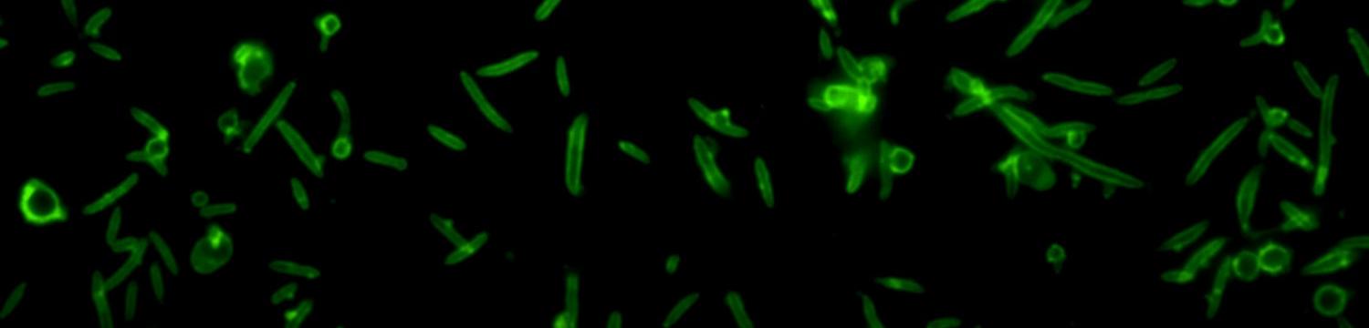
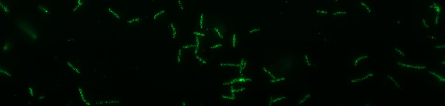
- Renzi, F, Ittig, SJ, Sadovskaya, I, Hess, E, Lauber, F, Dol, M, Shin, H, Mally, M, Fiechter, C, Sauder, U, Chami, M & Cornelis, G 2016, Evidence for a LOS and a capsular polysaccharide in Capnocytophaga canimorsus Scientific Reports. https://doi.org/10.1038/srep38914

- Renzi F., Zähringer U., Chandler C.E., Ernst R.K., Cornelis G.R., Ittig S.J. Modification of the 1-phosphate group during biosynthesis of Capnocytophaga canimorsus lipid A. Infection and Immunity. 2015 Dec 7. pii: IAI.01006-15.

- Zähringer U., Ittig S., Lindner B., Moll H., Schombel U., Gisch N., Cornelis G.R. NMR-based structural analysis of the complete rough-type lipopolysaccharide isolated from Capnocytophaga canimorsus. J Biol Chem. 2014 Aug 22;289(34):23963-76. doi: 10.1074/jbc.M114.571489. Epub 2014 Jul 2.

- Ittig, S., Lindner, B., Stenta, M., Manfredi, P., Zdorovenko, E., Knirel, Y., Dal Peraro, M., Cornelis, G.R, Zähringer, U. The Lipopolysaccharide from Capnocytophaga canimorsus Reveals an Unexpected Role of the Core-Oligosaccharide in MD-2 Binding. PLoS pathogens 2012, 8(5), e100266

- Shin H., Mally M., Meyer S., Fiechter C.,Paroz C., Zähringer U., Cornelis G.R. Resistance of Capnocytophaga canimorsus to killing by human complement and polymorphonuclear leukocytes. Infection and Immunity 2009, 77(6): 2262–2271. Epub Mar 23, 2009


Interplay between C. canimorsus and the coagulation system

C. canimorsus sepsis is accompanied in up to 50% of the cases by skin manifestations, reaching from petechiae to purpura fulminans. The patients that develop severe sepsis and septic shock frequently suffer from disseminated intravascular coagulation (DIC) and consequently develop gangrene (8-13% of all cases) leading to amputations of fingers, arms or legs. One third of the autopsies carried out on deceased patients showed haemorrhage of the adrenal glands. Haemostasis is part of the innate immunity and a range of bacteria has the capacity to modulate this mechanism of defence. In view of these facts and the frequent occurrence of bleeding abnormalities during C. canimorsus infections we investigate the interactions between C. canimorsus and the human coagulation system.  This research is done in collaboration with the group of JM Dogné from the "Namur Medicine & Drug Innovation Center" (NAMEDIC).

Publications:

Hack, K, Renzi, F, Hess, E, Lauber, F, Douxfils, J, Dogné, JM & Cornelis, GR 2016, Inactivation of human coagulation factor X by a protease of the pathogen Capnocytophaga canimorsus Journal of thrombosis and haemostasis : JTH. DOI: 10.1111/jth.13605


Polysaccharide Utilization Loci (PUL) of C. canimorsus

A hallmark of Bacteroidetes is their remarkable capacity to harvest and metabolize all kinds of complex carbohydrates.  This property depends on specialized molecular complexes that are assembled at their surface.  The archetype of these complexes is the Starch Utilization System (Sus) of Bacteroides thetaiotaomicron.  These complexes are encoded in genomic loci called Polysaccharide Utilisation Loci (PUL). The genome of our reference strain C. canimorsus 5 contains13 PUL and these encode more than half of all proteins exposed at the bacterial surface.  We have discovered that PUL5 encodes the Glycoproteins Deglycosylation complex (Gpd) enabling the bacterium to harvest amino sugars from the surface of eukaryotic cells including phagocytes as well as from soluble serum proteins, such as IgG and transferrin.  The system is essential during the human infection because it represents for C. canimorsus the only source of amino sugars necessary for the synthesis of their peptidoglycan. We also discovered that PUL3 encodes a novel iron acquisition system allowing the bacterium to fetch iron from human transferrin. Finally, PUL9 encodes the Muc complex, devoted to mucin degradation, an abundant carbohydrate source for C. canimorsus in its ecological niche, the dog’s mouth.


Main publications:

- Renzi F., Manfredi P., Dol M., Fu J., Vincent S., Cornelis G.R. Glycan-foraging systems reveal the adaptation of Capnocytophaga canimorsus to the dog mouth. MBio. 2015 Mar 3;6(2):e02507. doi: 10.1128/mBio.02507-14.

- Manfredi, P., Lauber, F., Renzi, F., Hack, K., Hess, E. & Cornelis, G. R. New iron acquisition system in Bacteroidetes. Infection and Immunity. 83, 1, p. 300-310 11 p.

- Manfredi P., Renzi F., Mally M., Sauteur L., Schmaler M., Moes S., Jenö P., Cornelis G.R.  The genome and surface proteome of Capnocytophaga canimorsus reveal a key role of glycan foraging systems in host glycoproteins deglycosylation. Mol Microbiol. 2011 Aug;81(4):1050-60. doi:10.1111/j.1365-2958.2011.07750.x.

- Renzi F., Manfredi P., Mally M., Moes S., Jenö P., Cornelis G.R. The N-glycan Glycoprotein Deglycosylation Complex (Gpd) from Capnocytophaga canimorsus Deglycosylates Human IgG. PLoS Pathog. 2011 Jun;7(6):e1002118.

- Mally M., Shin H., Paroz C., Landmann R., Cornelis G.R.: Capnocytophaga canimorsus: a human pathogen feeding at the surface of epithelial cells and phagocytes. PloS Pathog. 2008 Sep 26;4(9):e1000164. doi: 10.1371/journal.ppat.1000164.


How are lipoproteins exported to the bacterial surface?

Lipoproteins represent the biggest part of the PUL-encoded complexes and they are surface-exposed. This abundance of exposed lipoproteins is one of the main characteristics of Bacteroidetes. In Proteobacteria, the vast majority of outer membrane lipoproteins is inserted in the inner leaflet of the outer membrane and thus faces the periplasm.

Starting from the hypothesis that in Bacteroidetes surface exposed lipoproteins are endowed with a specific signal, we focused at identifying and characterizing this putative signal. In order to see if a specific amino acid motif would be responsible for the targeting of lipoproteins to the bacterial surface, we performed an in silico analysis on the sequences of the surface exposed lipoproteins of C. canimorsus. This led to the discovery of a consensus motif (QKDDE), located at the N-terminus of these proteins. In order to see whether this motif would represent an export signal, we grafted it to sialidase (SiaC), an OM-anchored periplasmic lipoprotein, and monitored its localization by immunofluorescence (IF) using both flow cytometry and microscopy. We observed that modified SiaC was exposed to the surface while normal SiaC was not. This showed that the consensus motif is indeed the lipoprotein export signal (LES) of C. canimorsus. We then analysed the motif in detail and found that the minimal LES consists of C-X-K-(D/E)2-X with the constraint that the overall charge must be negative (Fig.1). We also performed a similar in silico analysis in two other species of the phylum Bacteroidetes, namely  B.acteroides fragilis and Flavobacterium johnsoniae, and identified the SDDDD and SDDFE consensus motifs respectively. Both motifs were grafted to SiaC and found to be sufficient to drive the protein to the surface. Taken together, these results show that the LES identified are quite conserved and confirm the existence of a shared novel pathway for lipoprotein export in this phylum of Gram-negative bacteria .Our current work focuses on unravelling this new pathway…


Publications:

- Lauber, F, Cornelis, G & Renzi, F# 2016, Identification of a New Lipoprotein Export Signal in Gram-Negative Bacteria mBio. DOI: 10.1128/mBio.01232-16 # corresponding author